


MIT xvr speeds patient-specific X-ray guidance for surgery
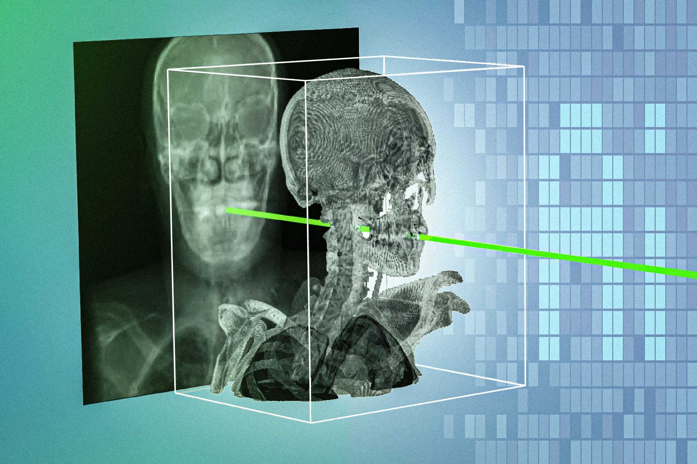
Researchers at MIT, working with clinicians and collaborating institutions, have developed xvr, an artificial intelligence technique that aligns X-rays taken during minimally invasive surgery with a patient’s preoperative CT or MRI scan. The system adapts to a new patient in about five minutes, performs the matching in seconds, and achieves sub-millimeter precision.
The method, short for X-ray volume registration, outperformed existing AI approaches by an order of magnitude across a broad range of patients, body parts, and procedures. The research paper, “Rapid patient-specific neural networks for X-ray to volume registration,” has been published in Nature.
Turning 2D X-rays into more useful navigation
Clinicians use real-time X-rays to guide catheters, endoscopes, and other instruments through small incisions in procedures such as angioplasty. Yet an X-ray is a flat image, making it difficult to establish the exact location and orientation of a tool relative to the patient’s anatomy. Registration with a preoperative three-dimensional scan can supply that context.
Current manual registration can be slow and demanding. It may require clinicians to enter positioning values or select anatomical landmarks on a screen. AI models intended to automate the task have also faced a central problem: anatomy varies substantially between patients, while high-quality annotated image data are limited.
Xvr takes a different approach. Rather than relying on one model intended to work equally well for everyone, it creates a model tailored to the individual patient. Vivek Gopalakrishnan, lead author and a postdoctoral researcher in MIT’s Computer Science and Artificial Intelligence Laboratory, said the aim is to make 2D X-rays more informative so procedures can be safer and easier to perform.
Physics-based synthetic images support fast adaptation
Starting from a patient’s CT or MRI, xvr produces thousands of synthetic X-rays from multiple angles at a rate of about 1,000 images per second. A physics-based simulation of the X-ray process is used to make the generated images realistic. Because the simulation is based entirely on that patient’s scan, the researchers say there is no room for hallucinations in the image-generation stage.
Training a patient-specific registration model from scratch would take roughly 12 hours, which is impractical for emergency use. To reduce that time, the team used xvr to pretrain a foundation model on whole-body 3D scans from more than 2,000 patients. The scans covered different ages, imaging modalities, and anatomical regions, allowing the pretrained system to adapt rapidly to a new patient while retaining the accuracy of a model trained from scratch.
Validation and deployment remain the next steps
The researchers evaluated xvr using the largest available dataset of real 2D/3D registrations. The dataset included material from five hospitals and covered dozens of bones and organ systems in adult and pediatric patients. The model was accurate and robust compared with other AI-based methods, while operating quickly enough for emergency surgery scenarios described by the team.
The work may also improve surgical robotics technologies. The researchers plan further reliability studies, work to support real-time deployment, and extensions for more complex cases such as moving body parts. For healthcare providers and surgical-technology teams, the practical implication is to assess patient-specific 2D/3D registration as a potential navigation component, while keeping clinical validation and workflow integration central to any deployment decision.